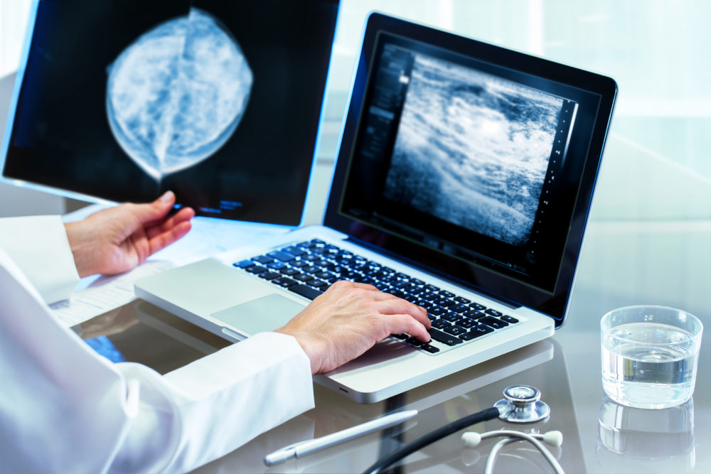
Dense breast tissue carries an increased risk of developing breast cancer. Because 40 percent of women have dense breast tissue, physicians have been looking for better ways to detect cancer among these women.
Mammograms have helped find cancer—and save lives—for many women. But the technology has limitations, particularly in screening dense breast where cancer can be harder to spot. On a mammogram, dense tissue is white and fat tissue is gray. Doctors look for white lumps, which could be cancer. These white lumps are difficult to see on a white background of dense breast tissue. It is like looking for a white balloon in a white cloud.
The screening process is different from a mammogram. Women lie down for the test and a technologist scans the breast with a gel-covered paddle. The scan records up to a thousand images for the radiologist to review.
“40 percent of women have dense breast tissue, physicians have been looking for better ways to detect cancer among these women”
“The radiologist can examine all the data and look at the breasts in multiple planes,” said Elizabeth Jekot, MD, medical director of the center. “This technology is a game-changer for screening dense-breasted women, allowing for earlier detection and maximizing survival.”
Dr. Jekot says that adding ABUS to screening mammography finds 55 percent more breast cancers than mammography alone in women with dense breasts.
The new technology uses three dimensional (3-D) imaging, which is the key to detecting tumors. Two-dimensional imaging with ultrasound was the standard, but new 3-D imaging allows for more sensitivity in finding tumors. The test takes 15 minutes per breast and there is very little compression. The system is designed to enhance the consistency, reproducibility and sensitivity of whole breast ultrasound, demonstrating improvement in cancer detection in women with dense breasts without prior breast intervention.
In 2011, the Texas Legislature passed House Bill 2102, known as “Henda’s Law”?named after Henda Salmeron, a Texas real estate agent and breast cancer survivor who was instrumental in organizing the effort to pass the law in Texas. Based on a similar law in Connecticut, HB 2102 requires that mammography centers inform women that dense breast tissue can affect the accuracy of mammography in detecting breast cancer. These identified women may benefit from supplemental breast screening.
For more information visit Baylor Scott & White.


Recent Comments